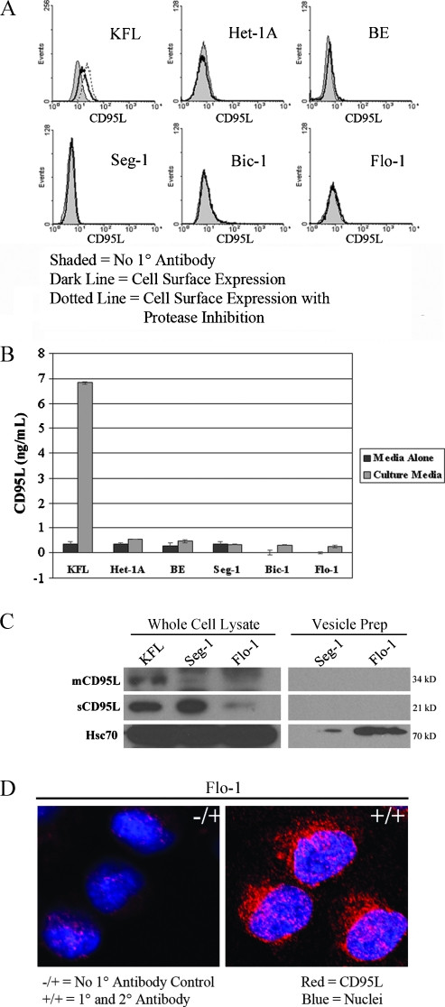
Figure 2.
Subcellular localization of CD95L. (A) FACScan analysis of CD95L cell surface expression (shaded histograms represent no primary antibody controls; open histograms represent experimental groups in the absence [solid lines] or presence [dashed lines] of MMP inhibition). Cell surface expression of CD95L was not detected in any of the esophageal or adenocarcinoma cell lines, even after MMP inhibition. (B) CD95L ELISA of culture medium. Data are presented as mean concentration (ng/ml) ± SEM of three individual experiments. Secretion of CD95L by the esophageal and adenocarcinoma cell lines was negligible or absent. (C) Immunoblot of CD95L expression in whole cell lysates and vesicle preparations from the adenocarcinoma cell lines Seg-1 and Flo-1. Vesicles were detected and isolated from Flo-1 cells and to a lesser degree from Seg-1 cells (as evidenced by Hsc70 immunoreactivity [36]), but CD95L was not detected by immunoblot analysis these vesicle preparations. (D) Laser scanning confocal microscopy of CD95L in the EA cell line Flo-1 (magnification, x400). CD95L is predominantly localized to the cytoplasm in a perinuclear distribution. -/+ indicates represents no primary antibody control; blue, nuclei; red, CD95L. All results are representative of at least three individual experiments.

