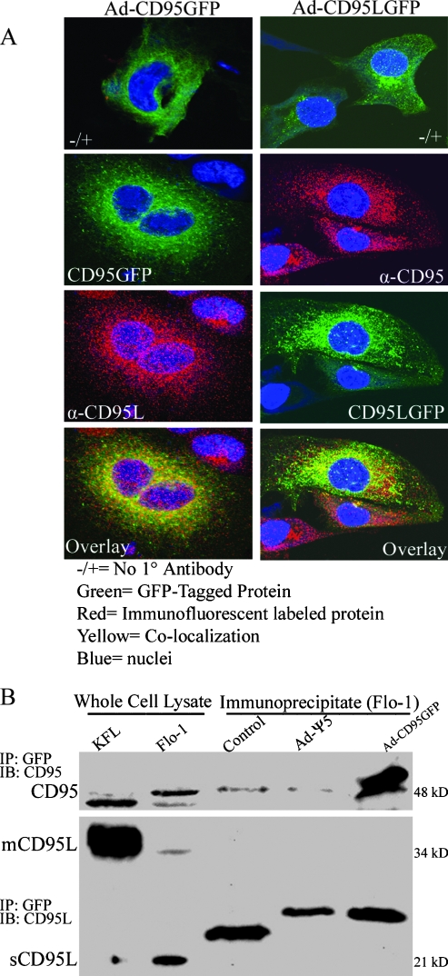
Figure 3.
Intracellular interaction of CD95 and CD95L. (A) Laser scanning confocal microscopy of CD95 and CD95L in Flo-1 cells after infection with either Ad-CD95-GFP (left column) or Ad-CD95L-GFP (right column) as described in Materials and Methods (magnification, x400). In the left column, CD95 appears green and CD95L appears red, whereas CD95 appears red and CD95L appears green in the right column. Similar to the expression of the native proteins, both Ad-CD95-GFP and Ad-CD95L-GFP predominantly localize in the cytoplasm. A modest degree of CD95-CD95L colocalization was apparent after infection with both adenoviral constructs. (B) Immunoblot of CD95 and CD95L in Flo-1 cells after infection with Ad-CD95-GFP and immunoprecipitation of GFP. GFP immunoprecipitation was confirmed by detection of CD95 at an expected, slightly larger molecular weight, but neither mCD95L nor sCD95L was coprecipitated. Nonspecific immunoprecipitate bands in the CD95L immunoblot represent BSA antibody light chain (Control) and GFP antibody light chain (Ad-Ψ5 and Ad-CD95-GFP). Results are representative of three individual experiments.

