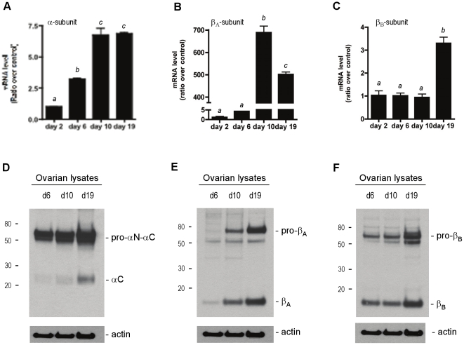
Figure 1. Expression of inhibin α-, βA- and βB-subunits in the postnatal mouse ovary.
Basal expression levels of inhibin α- (A), βA- (B) and βB- (C) subunits in developing mouse ovaries were determined by real-time RT-PCR. The mRNA levels of the subunits are shown as a ratio over the day 2 ovary mRNA levels (control). Detection of the inhibin α- (D), βA- (E) and βB- (F) subunit protein in ovarian lysates from day 6, day 10 and day 19 ovaries. Ovarian lysates were collected, separated under reducing conditions; each subunit was detected by immunoblotting with the corresponding rabbit polyclonal antibody. Forty µg of protein was loaded per lane. Equal loading of lysates was confirmed with an anti-actin antibody.

