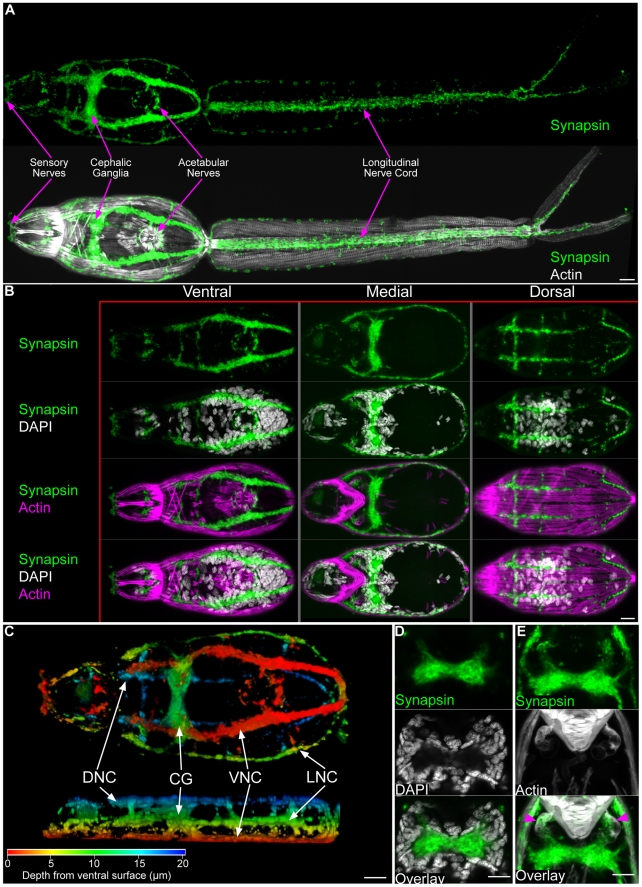
Figure 4. The central nervous system of cercariae.
(A) Immunofluorescence with an anti-synapsin antibody that labels the cephalic ganglia and many peripheral neural structures. Below, synapsin labeling is shown together with phalloidin staining. (B) Maximum confocal projections generated from ventral, medial, or dorsal focal planes. Anatomical regions are given above and staining reagents are listed to the left. (C) Depth projection showing synapsin staining in the CNS. Ventral view is shown above and a lateral view is shown below. Scale shown below indicates color-coding of distances from the ventral surface. Abbreviations: Dorsal nerve cords (DNC), cephalic ganglia (CG), ventral nerve cords (VNC), lateral nerve cords (LNC). For the sake of simplicity we have not distinguished anteriorly projecting cords from dorsally projecting cords. (D) Single confocal section though the cephalic ganglia, showing the central neuropil surrounded by neuronal nuclei. (E) Confocal projection showing innervation between the cephalic ganglia and the musculature surrounding the anterior ducts of the acetabular glands (magenta arrowheads). Scale bars, 10 µm. Anterior faces left in panels A, B, and C and up in panels D and E.

