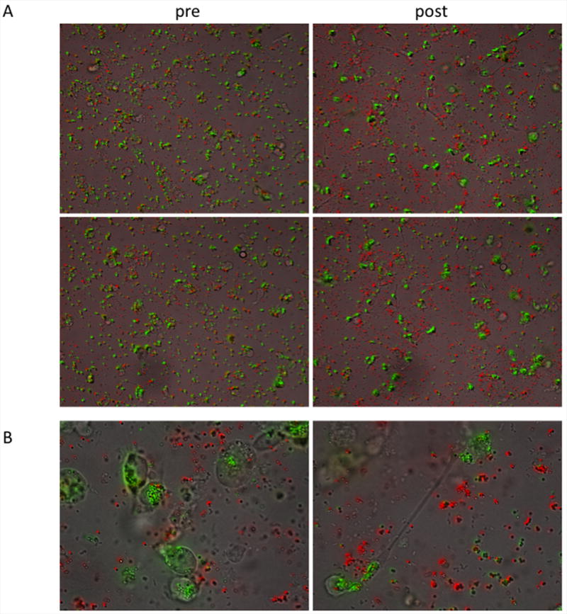
Figure 9. Time-lapse microscopy of phagocytosis.

Antibody-coated (green) and bare (red) beads were incubated with THP-1 cells on coverslips for time lapse microscopy, demonstrating antibody-specific uptake. A. 20× magnification still images representing the first (left) and final (right) frames from two 14 hour experiments. B. Representative 63× magnification images at 14 hours.
