
Figure 1. Modification of tubulin cysteines by HOCl and chloramines.
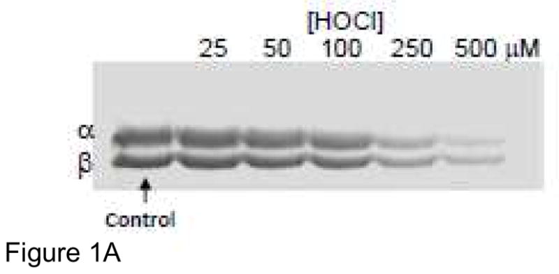

A) Tubulin samples (3 μM, 60 μM cys) were treated with 25, 50, 100, 250 or 500 μM HOCl for 10 min. at RT. Samples were incubated with 1.5 mM IAF for 30 min at 37 °C and then subjected to SDS-PAGE under reducing conditions on 7.5% polyacrylamide gels. Gel images were captured using a UV transilluminator and a Kodak DC290 imaging system. B) Tubulin samples (25 μM, 500 μM cys) were treated with 50, 100, 250 and 500 μM HOCl, GC or TC for 10 min at RT. Excess oxidant was scavenged with 1 mM met or monochlorodimedone (for HOCl). Samples were incubated with 2.5 mM IAF for 30 min at 37 °C and then protein was precipitated with 80% ethanol. Fluorescein-labeled protein pellets were collected, washed with 80% ethanol and resuspended in 3 M guanidine HCl in 0.1 M Tris pH 8.8. Fluorescein absorbance at 490 nm for standards and samples was measured in a 96 well plate. These data represent the mean +/− standard error of three independent experiments.