
Figure 4. Detection of interchain tubulin disulfides.
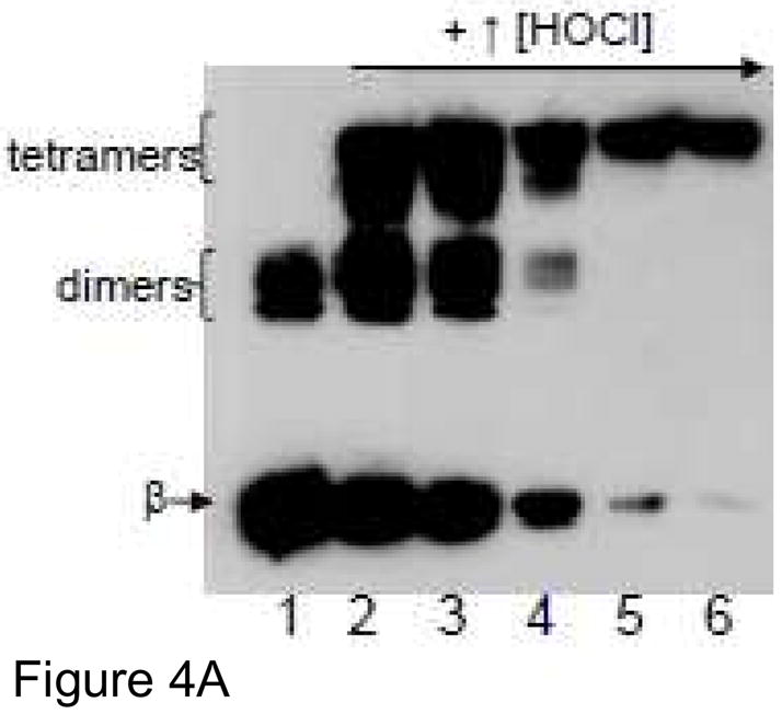
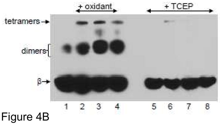
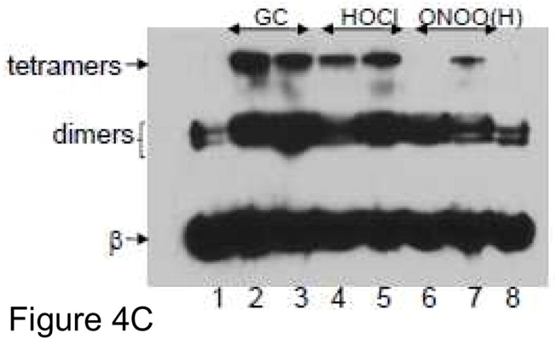
Oxidized tubulin species were separated by SDS-PAGE under nonreducing conditions on a 7.5% polyacrylamide gel and transferred to PVDF and probed with anti-β-tubulin. Monomeric β-tubulin (50 kDa) is labeled as well as dimers and tetramers. A) Tubulin samples (8 μM protein, 160 μM cysteines) were treated with 50, 100, 250, 500 and 1 mM HOCl for 10 min at 37 °C (Lanes 2–6). B) Tubulin (25 μM protein, 500 μM cys) was treated with 250 μM HOCl, GC or TC (lanes 2–4) for 10 min at RT. Identical oxidized tubulin samples were subsequently treated with 2.5 mM TCEP (lanes 6–8) for 5 min prior to electrophoresis. Control tubulin is in lanes 1 and 5 (+/− TCEP). C) Tubulin (25 μM protein, 500 μM cys) was treated with 100 or 250 μM GC (lanes 2–3), HOCl (lanes 4–5) and peroxynitrite (lanes 6–7) for 10 min at RT. Control tubulin is in lanes 1 and 8.