
Figure 5. Detection of methionine oxidation.
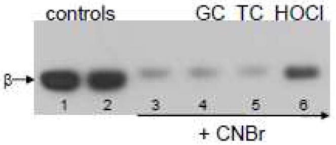
Tubulin samples (12.5 μM, 250 μM cysteines, 325 μM methionines) were treated with 250 μM of each oxidant for 30 min at RT. Samples were subsequently acidified with formic acid, and treated with CNBr for 16 hours. Samples were neutralized with NH4OH to pH 7.4–7.6 and subjected to SDS-PAGE under reducing conditions on a 7.5% polyacrylamide gel. Proteins were transferred to PVDF and probed with anti-β-tubulin. Lane 1 contains control tubulin, lane 2, control tubulin treated with formic acid and NH4OH, lane 3, tubulin treated with CNBr in the same manner as the oxidized tubulin samples in lanes 4–6.