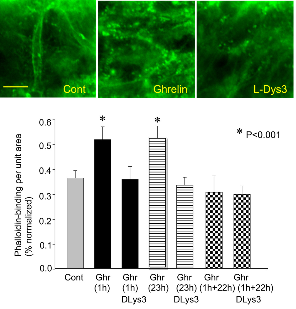
Figure 3.
Effect of ghrelin on phalloindin-binding. Photomicrographs show phalloidin-based fluorescence representing polymerized F-actin in dendrites and spines in control, ghrelin, and D-Lys-3-GHSR-6. The graph depicts phalloidin-binding in response to ghrelin-application for 1 hour and 23 hours. The slices were fixed at the end of ghrelin-application. Some slices were kept alive before fixation for 22 hours in the absence of ghrelin after 1 hour of ghrelin-incubation (labeled as 1h+22h; see text for details). The calibration in Cont (10 µm) is shared by all three photomicrographs. Cont: Control, Ghr: Ghrelin, DLys3: D-Lys3-GHSR-6. Asterisks indicate p<0.01.

