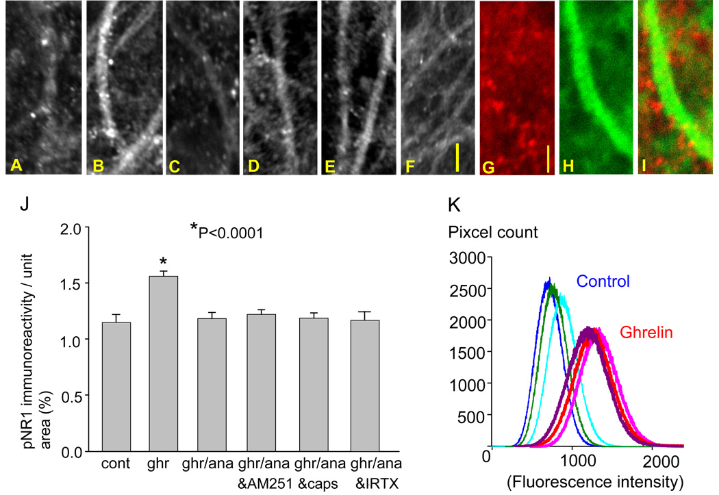
Figure 5.
Effects of ghrelin and R(+)-methanandamide on pNR1 expression. Photomicrographs show pNR1 immunoreactivity in the control (A), 200 nM ghrelin (B), 200 nM ghrelin and 20 nM R(+)-methanandamide (C), 200 nM ghrelin, 20 nM R(+)-methanandamide and 5 µM AM251 (D), 200 nM ghrelin, 20nM R(+)-methanandamide and 5 µM capsazepine (E), 200 nM ghrelin, 20nM R(+)-methanandamide and 10 nM iodoresiniferatoxin (IRTX)(F). Dual imaging of pNR1 (red) with phalloidin (green)(G, H, and I). The graph summarizes the results (J). The intensity of pNR1 immunofluorescence in the CA1 stratum radiatum was greater in the ghrelin-treated hippocampal slices compared with control slices (K). The calibration in F (10 µm) is shared by photo-micrographs in A–F, and the calibration in G (2 µm) is shared by photomicrographs in G–I.

