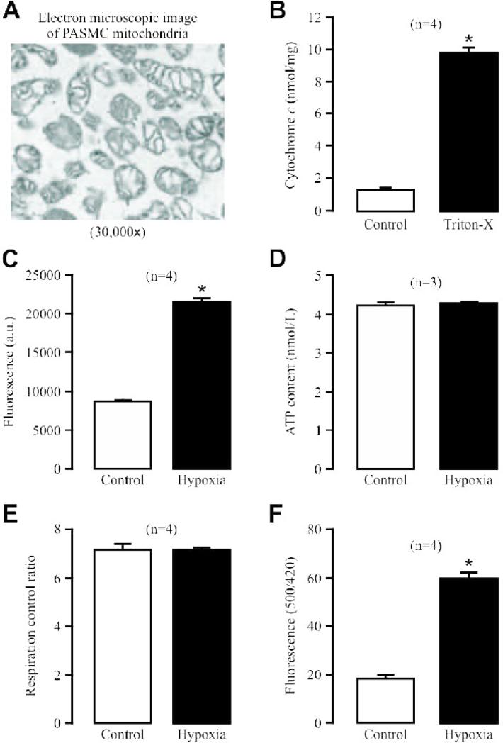
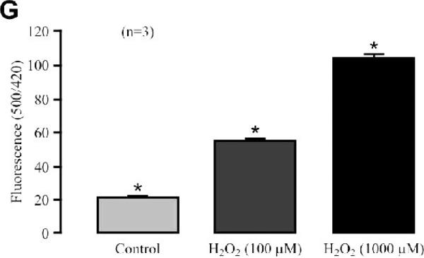
Fig. 1. Hypoxia causes a large increase in ROS production in isolated mitochondria from PASMCs.
(A) Representative electron microscopic image of isolated mitochondria. (B) Cytochrome c release in isolated mitochondria before and after application of Triton X-100 (1 mM) for 5 min. Numbers in parentheses mean the number of independent experiments performed. *P<0.05 compared with control (before application of Triton X-100). (C) Effect of acute hypoxic exposure for 5 min on ROS production, determined by assessing the fluorescence of the ROS detection dye H2DCF, in isolated mitochondria. (D) Effect of acute hypoxia on ATP production in isolated mitochondria. (E) Effect of hypoxia on respiration activity in isolated mitochondria. (F) Effect of hypoxia on mitochondrial inter-membrane space ROS production, examined by measuring the fluorescence of the specific ROS biosensor pHyPer, in isolated PASMCs. (G) Effect of H2O2 on HyPer signals in isolated PASMCs.


