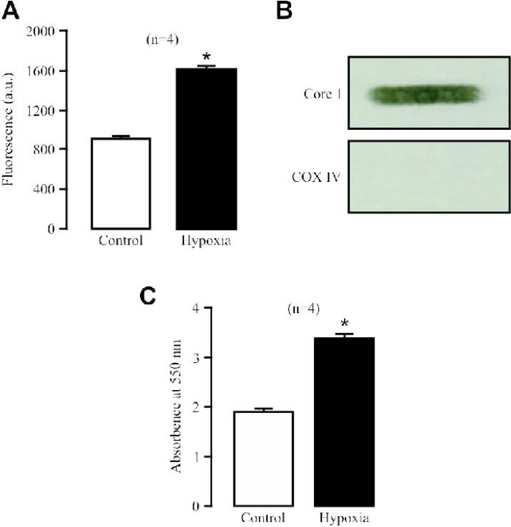
Fig. 2. Hypoxia significantly increases ROS production in isolated complex III from PASMCs.
(A) Effect of hypoxia on ROS formation (H2DCF fluorescence) in isolated complex III. (B) Representative Western blots show the presence of the complex III subunit Core 1 protein, but not the complex IV subunit COX IV protein in isolated complex III samples. (C) Effect of hypoxia on isolated complex III activity, gauged by assessing the absorbance of cytochrome c reduction at 550 nm.

