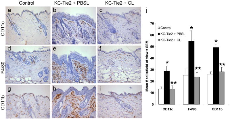
Figure 1. Clodronate liposome administration results in antigen cell depletion.

Immunohistochemical staining detecting CD11c+ (a-c) F4/80+ (d-f), and CD11b+ (g-i) cells in skin sections from KC-Tie2 or control animals treated with either clodronate or PBS filled liposomes for 6 weeks. Quantitation using interactive image analyses approaches (j) reveals increased numbers of antigen presenting cells in KC-Tie2 + PBS liposomes (PBSL) compared to control mice, and that these numbers return to control mouse levels following clodronate liposome (CL) administration. * p≤0.05 compared to controls; ** p≤0.05 compared to KC-Tie2 + PBSL.
