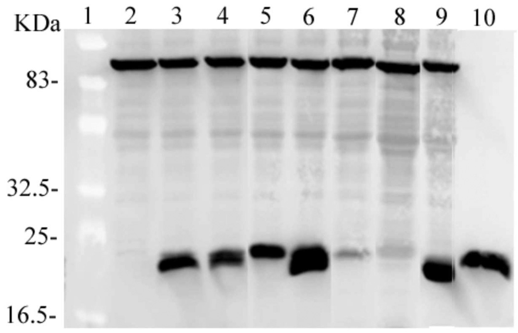
Figure 5. Expression of wild-type and mutant AGT proteins in E. coli TRG8.
Analysis by SDS-PAGE and western blot. Lane 1, molecular weight standards (10 µg total protein). Bands do not stain but can be seen as lighter zones against the background. Lane 2, proteins from TRG8 cells containing no plasmid (25 µl); lane 3, proteins from TRG8 cells containing pQE-hAGT-wild-type (25 µl); lane 4, proteins from cells containing pQE-hAGT-KDC(3–5)-AAA (25 µl); lane 5, proteins from cells containing pQE-hAGT-DCE(4–6)-AAA (25 µl); lane 6, proteins from cells containing pQE-hAGT-EWL(166–168)-AAA (25 µl); lane 7, proteins from cells containing pQE-hAGT-CEM(5–7)-AAA (25 µl); lane 8, AGT proteins from cells containing pQE-hAGT-VKE(164–166)-AAA (25 µl); lane 9, proteins from cells containing pQE-hAGT-KEW(165–167)-AAA (25 µl). Lane 10 contains wild-type AGT purified from XL1-blue E. coli cells (20 µg).

