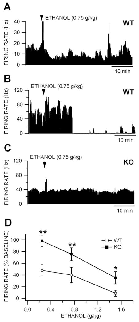
Figure 4. Inhibition of VTA GABA neuron firing rate is reduced in Cx36 KO mice.

(A) This ratemeter record shows a representative VTA GABA neuron in a WT mouse recorded in vivo with a baseline firing rate of approximately 15 Hz before and after IP administration of 0.75 g/kg ethanol. Intraperitoneal administration of 1.0 g/kg ethanol reduced this neuron’s activity ~50% at approximately 10 min after injection. Transient increases in firing rate are often associated with systemic administration of ethanol (Steffensen et al., 2009). (B) This ratemeter record shows a representative VTA GABA neuron recorded in a separate WT mouse with a baseline firing rate of approximately 40 Hz before and after IP administration of 0.75 g/kg ethanol. Intraperitoneal administration of 1.0 g/kg ethanol suppressed this neuron’s activity at approximately 10 min after injection. (C) The ratemeter record shows a representative VTA GABA neuron in a KO mouse, with a baseline firing rate that was similar to that of the neuron in the WT mouse in (B), before and after IP administration of 0.75 g/kg ethanol. Ethanol did not alter the firing rate of this neuron in a KO mouse. (D) This graph compares the effects of IP administration of 0.75-4.0 g/kg ethanol on the firing rate of VTA GABA neurons recorded in KO vs WT mice. Ethanol reduced firing rate in both WT and KO mice. However, the IC50 for ethanol inhibition was 0.25 g/kg in WT mice and 1.4 g/kg in KO mice. Wild-type mice were significantly more sensitive to ethanol than KO mice at all doses tested.
