
Fig. 3.
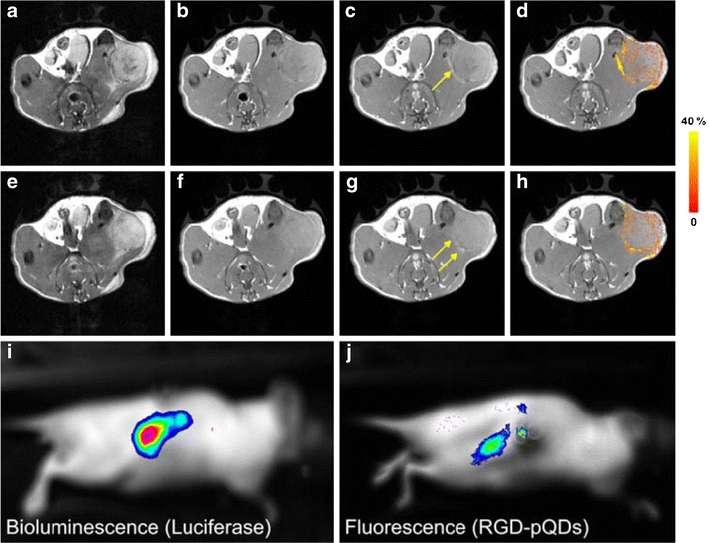
Magnetic resonance and optical molecular imaging of tumor angiogenesis using alpha-v-beta-3 targeted multimodal quantum dot-based nanoparticles. T2-weighted images (a, e), collected before the nanoparticles were injected, show the contour of the tumor on the flank. T1-weighted images (b, c, f, g) with TR = 800 ms were measured before (b, f) and 45 min after (c, g) the injection of the nanoparticles. The arrows in (c, g) indicate bright (positive contrast) regions in the periphery of the tumor. In d, h, pixels in the tumor with signal enhancement of at least three times the noise level are color coded according to the pseudo-color scale on the right. i, j Bioluminescence imaging and fluorescence imaging of a Balb/c nude mouse with a luciferase-expressing renal carcinoma tumor after injection of luciferin (i). The strong bioluminescence signal is indicative of tumor growth in the right kidney. This signal colocalizes with a strong fluorescence signal (j) originating from intravenously administrated nanoparticles that are accumulated in the tumor. Republished with kind permission from Mulder WJ, Castermans K, van Beijnum JR et al. Molecular imaging of tumor angiogenesis using alphavbeta3-integrin targeted multimodal quantum dots. Angiogenesis. 2009;12(1):17-24.