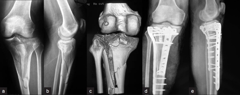
Figure 5.

Anteroposterior (a), lateral (b) radiographs, and three-dimensional CT scan (c) of Patient 4 shows the complex fractures of the tibial plateau and metaphysis. Postoperative anteroposterior (d) and lateral (e) X-ray films show the fractures were well reduced and fixed via combined PM and AL approaches. PM= Posteromedial, PL= Posterolateral
