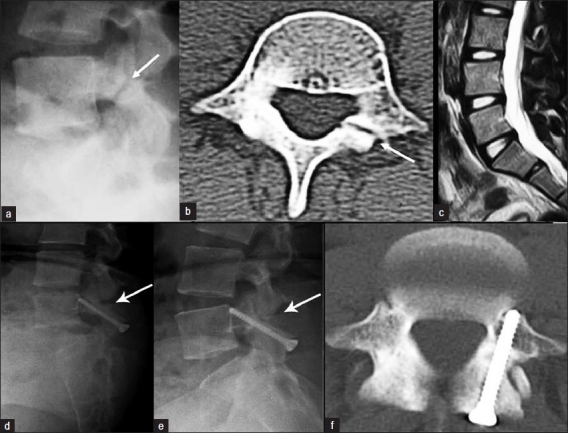
Figure 1.

(a) Preoperative lateral radiograph and (b) axial CT scan showing unilateral defect of the pars interarticularis of the L4 vertebra. (c) Sagittal T2 weighted MRI demonstrating the normal L4-L5 disc without any degeneration. Follow-up lateral dynamic radiographs in (d) flexion and (e) extension, showing complete healing of the defect without signs of instability. (f) Postoperative axial CT scan demonstrating complete healing of the spondylolytic defect
