
Figure 2.
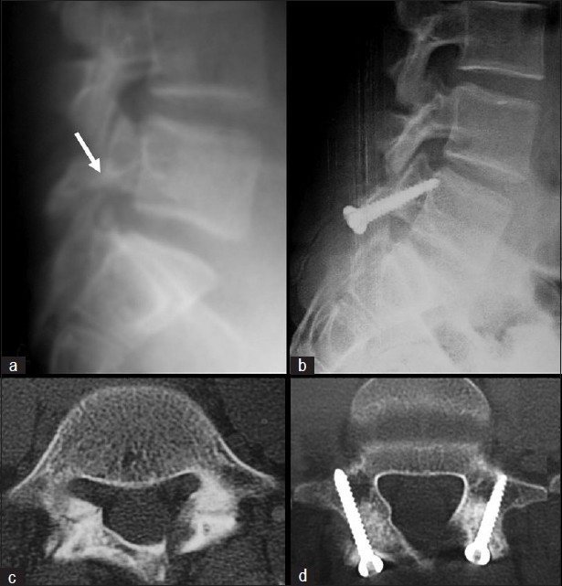
(a) Preoperative lateral radiograph and (b) follow-up lateral radiograph of a patient with bilateral spondylolysis of L5 fixed by Buck’s technique on both sides. (c) Preoperative and (d) follow-up axial CT scan of the same patient showing complete healing of the pars defect bilaterally, without signs of screw loosening, back out, or breakage