Figure 3.

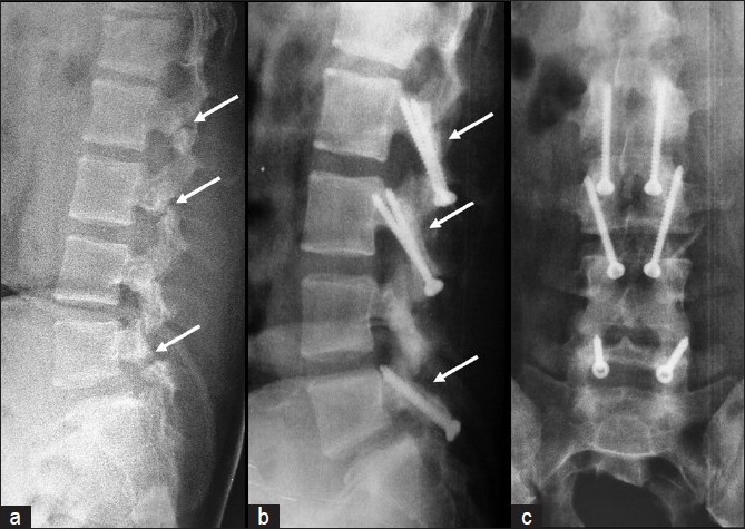
(a) Preoperative lateral radiograph of a patient with triple level spondylolysis at L2, L3, and L5. (b) Postoperative follow-up lateral and (c) anteroposterior radiographs showing good healing of the pars defect at all three levels

(a) Preoperative lateral radiograph of a patient with triple level spondylolysis at L2, L3, and L5. (b) Postoperative follow-up lateral and (c) anteroposterior radiographs showing good healing of the pars defect at all three levels