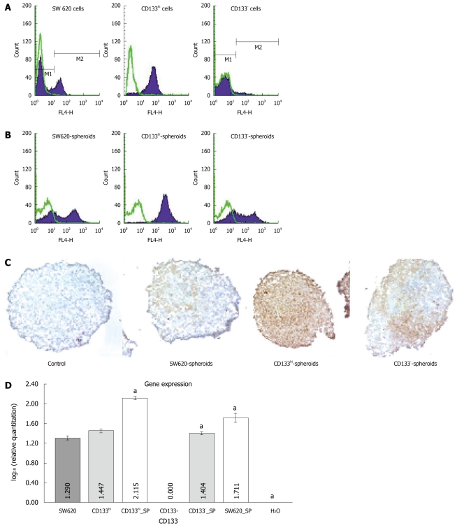
Figure 2.
Fluorescence-activated cell sorting showing CD133 expression in SW620, CD133- and CD133hi cells (A) and in their spheroids (B), CD133 staining in spheroids of SW620, CD133- and CD133hi cells (original magnification × 100, brown indicates positive staining) (C), and reverse transcription-polymerase chain reaction showing CD133 expression in SW620, CD133- and CD133hi cells and their spheroids. aP < 0.05 vs monolayer cells. SP: Spheroid.

