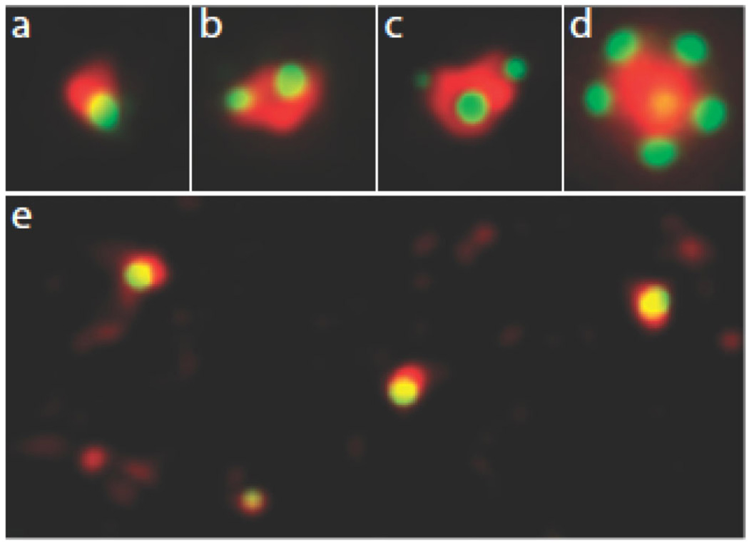
Figure 2. Fluorescence microscopic images of centrosomes in mammalian somatic cells.
The green contrast shows the localization of GFP-centrin 1, which is concentrated in the distal lumen of centrioles and thus serves as a centriole marker. The red contrast is gamma tubulin located in the pericentriolar material. (a) A single centriole associated with pericentriolar material during G1. (b) A centrosome in G2 containing a mother centriole (larger centrin dot) and a daughter centriole. The pair of centrioles is ofter referred to as a diplosome. (c) Spontaneous occurrence of two daughter centrioles with one mother centriole (a triplosome). This is seen at low frequency in some cell types such as CHO cells. (d) Overduplication of a mother centriole when Plk 4 is overexpressed. The mother centriole (seen weakly as the yellow dot in the middle of the gamma tubulin cloud) is driven to make more than one procentriole at one time giving a figure that resembles a rosette. (e) Multiple centrioles assembled de novo in a cell whose resident centrioles were ablated with a laser microbeam. Note the smaller size of the pericentriolar material and foci of gamma tubulin without centrioles.

