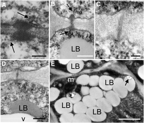
Figure 8.
Transmission Electron Micrographs of Dormant Meristems of Hybrid Aspen after Treatment with GA4.
(A) and (B) Examples of PD between L1 cells (A) and between corpus cells (B) in dormant meristems after 7 d treatment with water. The arrows point to a strand of endoplasmic reticulum at the PD.
(C) and (D) Callose-containing sphincters were no longer visible at PD after 5 d of GA feeding. Examples of PD between corpus cells (C) and rib meristem cells (D) resembling those in actively growing meristems. LBs were typically merging into vacuoles (D).
(E) At an earlier time point of 3 d, LBs are still present, but LB–cell wall connections were scarce.
Bars = 200 nm in (A) to (C), 100 nm in (D), and 1 μm in (E). For overviews, controls, and details, see Supplemental Figure 5 online.

