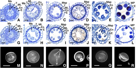
Figure 4.
Comparison of Male Gametogenesis in the Wild Type and the pss1 Mutant.
(A) to (F) and (M) to (O) show the wild type; (G) to (L) and (P) to (R) show the pss1 mutant. The cross sections ([A] to [L]) are stained with 0.25% toluidine blue O. E, epidermis; En, endothecium; ML, middle layer; Ms, microsporocyte; Msp, microspore; MP, mature pollen; S, sperm nuclei; T, tapetum; V, vegetative nuclei. Bars = 15 μm.
(A) and (G) Cross section of single locule at the microspore mother cell stage.
(B) and (H) Cross section of single locule at the meiosis stage.
(C) and (I) Cross section of single locule at the young microspore stage.
(D) and (J) Cross section of single locule at the vacuolated pollen stage.
(E) and (K) Cross section of single locule at the pollen mitosis stage showing two types of pollen grains in the mutant locule.
(F) and (L) Cross section of single locule at the mature pollen stage showing two types of pollen grains in the mutant locule.
(M) and (P) DAPI staining of a uninucleate stage microspore.
(N) and (Q) DAPI staining of a bicellular stage microspore.
(O) and (R) DAPI staining of a tricellular stage microspore.

