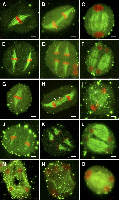
Figure 8.
Immunostaining of Spindles in Wild-Type and pss1 Male Meiocytes.
Chromosomes are stained by propidium iodide (red), and microtubules are labeled by fluorescein isothiocyanate (green). Bars = 5 μm.
(A) to (F) Male meiocytes of the wild type.
(A) Bipolar-oriented spindle at metaphase I.
(B) Two sets of univalents separated by the spindle at anaphase I.
(C) Phragmoplast starts to form between the two daughter pronuclei at telophase I. The weak staining in the midzone indicates the formation of a cytokinetic plane.
(D) Two sets of univalents aligned at the metaphase II plates.
(E) Two sets of univalents are separated to the opposite poles by the anaphase II spindles.
(F) At telophase II, new phragmoplast structure forms near the equatorial plate.
(G) to (O) Male meiocytes of the pss1 mutant.
(G) At metaphase I, the pss1 mutant shows normal-shaped spindles, but one or several chromosomes are delayed (indicated by arrowhead).
(H) Bivalents are pulled by the forces of anaphase I spindles and move away from the equator. Delayed chromosomes are observed (indicated by arrowheads).
(I) The telophase I spindle is somewhat disorganized compared with that of the wild type. In addition, delayed chromosomes are observed (indicated by arrowheads).
(J) The metaphase II spindle has a normal shape but is thinner than that of the wild type. Delayed chromosomes can also be seen (indicated by arrowhead).
(K) The shape and density of the anaphase II spindles are similar to that of the wild type.
(L) Most meiocytes of pss1 mutant show normal shaped spindles at telophase II.
(M) to (O) A small portion of meiocytes has disorganized microtubules and form atypical tetrads.

