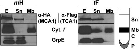
Figure 1.
MCA1 and TCA1 Are Soluble Proteins.
Cell extracts (E) from tF and mH strains, overlaid on a 1.5 M sucrose cushion, were separated by ultracentrifugation into the fractions schematically depicted in the right panel. Sn, supernatant; Mb, membrane; C, cushion; P, pellet. Equal volumes of each fraction were immunoreacted with anti-HA or anti-Flag antibodies. In addition, GrpE and cytochrome f were used as markers of soluble and membrane fractions, respectively. In the tF (TCA1) panel, fraction Sn was slightly contaminated by membranes, which explains the presence of cytochrome f (Cyt. f). Fractions C and P are not shown, as they were devoid of significant amounts of MCA1, TCA1, GrpE, or cytochrome f.

