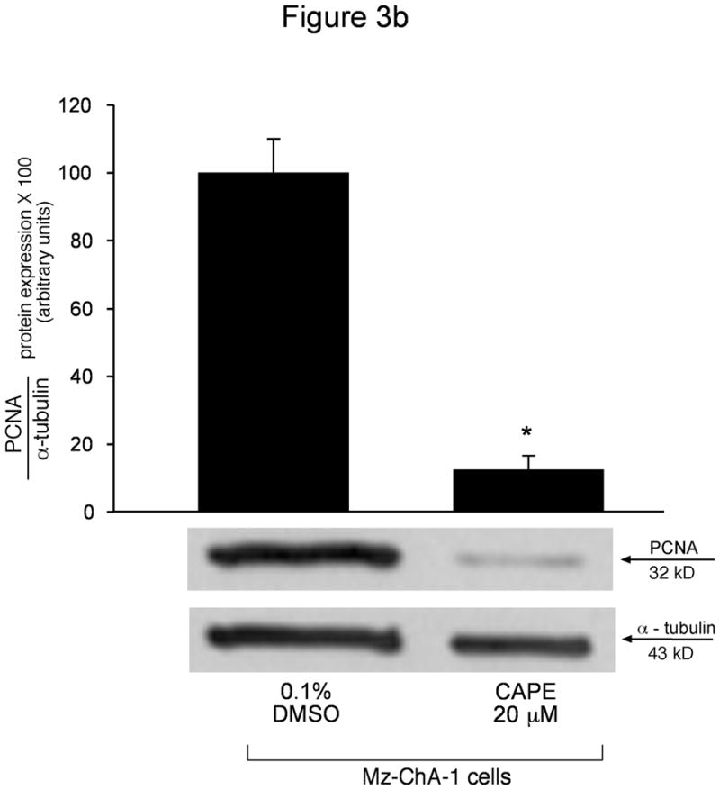
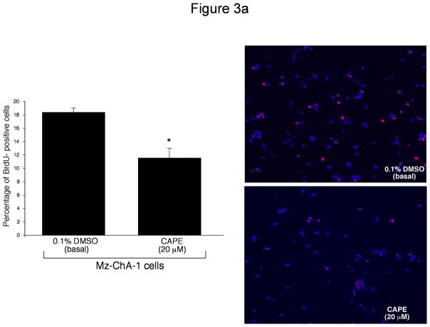
Figure 3.

[a] BrdU incorporation for measuring cell cycle progression in Mz-ChA-1 cells stimulated with 0.1% DMSO (basal) or CAPE (20 μM with 0.1% DMSO) for 48 hours demonstrated that in basal conditions the number of BrdU positive cells was approximately 50%. However, when Mz-ChA-1 cells were stimulated with CAPE, the number of BrdU positive cells decreased to approximately 10% thus decreasing cell cycle progression. The number of BrdU-positive nuclei were counted and expressed as a percentage of total cells in 5 random fields for each treatment group. *p<0.01 compared to basal (0.1% DMSO). [b] PCNA protein expression is markedly decreased by CAPE treatment (20 μM for 48 hours) compared to basal (0.1% DMSO). Alpha-tubulin levels were unchanged between basal- and CAPE- stimulated cell lysates. Data are ± SEM of 6 experiments. *p < 0.05 compared to basal (0.1% DMSO). A representative blot is shown.

