Figure 1.

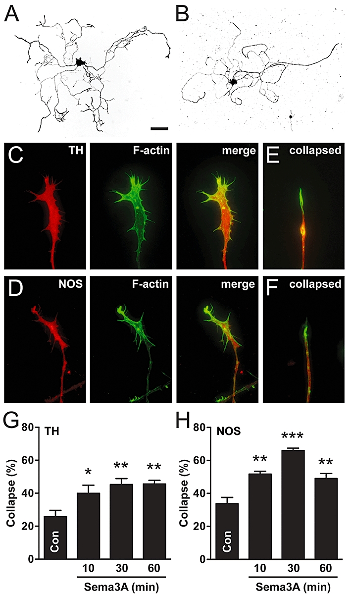
Sema3A causes growth cone collapse in adult sympathetic and parasympathetic neurones. (A,B) Inverted fluorescence images of cultured sympathetic (A; TH-positive) and parasympathetic (B; NOS-positive) pelvic ganglion neurones. (C, D) Intact growth cones of sympathetic (C) and parasympathetic (D) neurones. (E, F) Collapsed growth cones of sympathetic (E) and parasympathetic (F) neurones (merged images of TH or NOS with actin staining). Note the retraction of F-actin rich filopodia in collapsed growth cones. (G,H) Sema3A increased growth cone collapse in both sympathetic and parasympathetic neurones, at all time points tested (n= 4). Dunnett's test: *P < 0.05, **P < 0.01, ***P < 0.001 versus control. Scale bar = 100 µm in (A and B), 12 µm in (C–F). NOS, nitric oxide synthase; TH, tyrosine hydroxylase.
