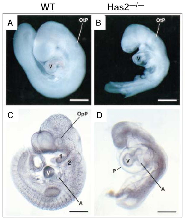
Figure 3. HAS2-deficient mice.
Disruption of hyaluronan synthase 2 resulted in the defect in heart formation. Representative wild-type (A) and Has2−/− (B) embryos at E9.5. Note the diminished size, the bloodless heart, and distorted somites of the Has2−/− embryo. E9.5 wild-type (C) and Has2−/− (D) embryos stained for the endothelial marker PECAM. Note the absence of an organized vascular network expressing PECAM in the Has2−/− embryo. P, pericardium; E, endoderm; M, mesoderm; OpP, optic placode; OtP, otic placode; first and secondpharyngeal pouches are numbered. From Camenisch et al., J. Clin. Invest. 106:349–360 (2000), with permission.

