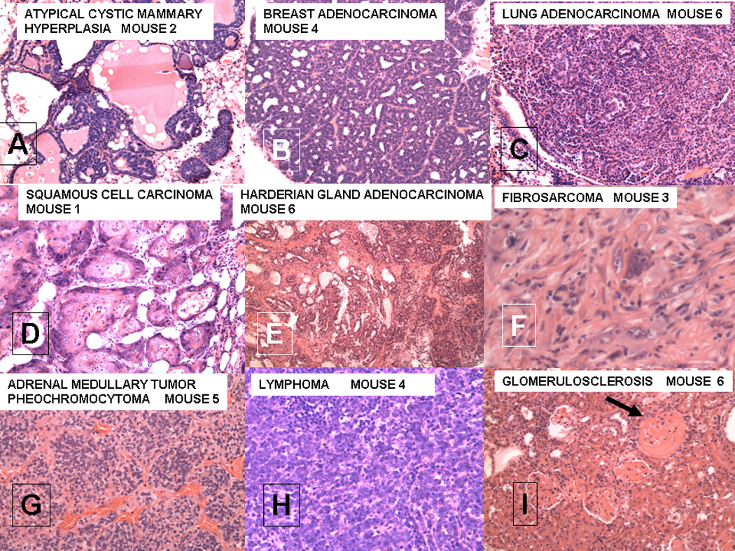
Figure 1.


A–I. Various lesions seen in female FVB/N mice after lethal irradiation and bone marrow transplantation from male MMTV-PyMT donors;
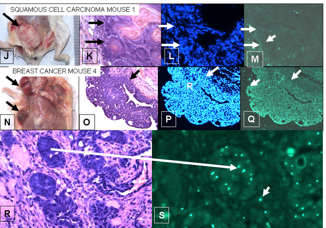
J–M. Squamous cell carcinoma from mouse 1;
N–S. Breast cancer from mouse 4.
For A–I, each section is stained with hematoxalin and eosin. All magnifications are 20X except for F which is 40X. The microscopic patterns of the breast adenocarcinoma were varied with medullary, papillary and glandular forms.
J–M Squamous cell carcinoma from mouse 1. The squamous cell carcinoma in the breast of mouse 1 does not contain Y-Chromosomes, whereas the breast adenocarcinoma in mouse 4 (E–F) does. J. Gross lesion from mouse 1. K. H&E, L. DAPI (nuclei), M. in situ for Y-chromosome (40X). The arrows mark the border between the squamous cell carcinoma and the stroma. D shows strong Y-chromosome staining in the stroma of the tumor, but the tumor cells are negative.
N–S. Breast Cancer from mouse 4. N. Gross lesions in breast of mouse 4. The upper arrow point to a cystic lesion; the lower to a solid lesion. O. H&E showing a papillary structure. P. DAPI showing nuclei in the tumor. Q. In situ for Y-chromosomes (20X) showing up to 80% of nuclei in tumor contain Y-chromosome in this section. The arrows in O-Q delineate the border of the stroma and the tumor. In contrast to M, Q shows that the tumor labels strongly for Y-chromosomes, whereas the stroma is weakly labeled. R. H&E and S in situ for Y-chromosome of breast adenocarcinoma (40X). The larger arrow shows a focus of cancer in the H&E section that is labeled for Y-chromosome in a serial section. Note the bright labeling of Y-chromosomes in the tumor. The smaller arrows in J point to Y-chromosome containing cells that could be either single tumor cells or blood derived mononuclear cells in the stroma.
