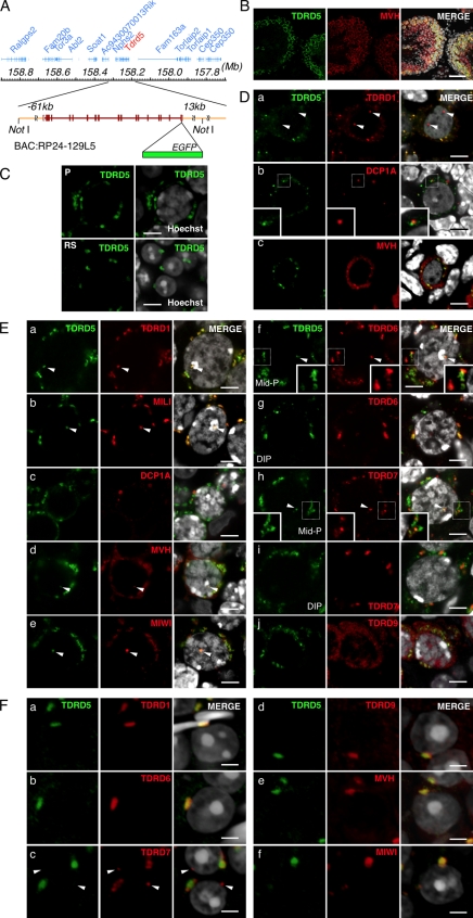
Figure 2.
TDRD5 localizes at IMCs and CBs in male germ cells. (A) The BAC bearing Tdrd5 and surrounding genes. EGFP was recombined in front of the stop codon of Tdrd5. (B) TDRD5-EGFP in an adult testis section costained with MVH and DAPI. The right shows merged images with Hoechst (from B to F). (C) TDRD5-EGFP in a pachytene spermatocyte (P) and round spermatids (RS). (D) Localization of TDRD5-EGFP (green) costained with TDRD1 (a), DCP1A (b), and MVH (c; red) in prospermatogonia at E17.5. Arrowheads in a indicate the nuclear localization of TDRD1. Insets in b show magnified views of the boxed regions. (E) Localization of TDRD5-EGFP (green) costained with TDRD1 (a), MILI (b), DCP1A (c), MVH (d), MIWI (e), TDRD6 (f), TDRD7 (h), and TDRD9 (j; red) in pachytene spermatocytes or with TDRD6 (g) and TDRD7 (I; red) in diplotene spermatocytes. Arrowheads indicate TDRD5-positive nuclear foci. Insets in f and h show magnified views of the boxed regions. (F) Localization of TDRD5-EGFP (green) costained with TDRD1 (a), TDRD6 (b), TDRD7 (c), TDRD9 (d), MVH (e), and MIWI (f; red) in round spermatids in the stage I seminiferous tubules. Arrowheads in c indicate the lone TDRD7-positive spot. Bars: (B) 50 µm; (C, D, and E) 5 µm; (F) 3 µm.

