Figure 6.
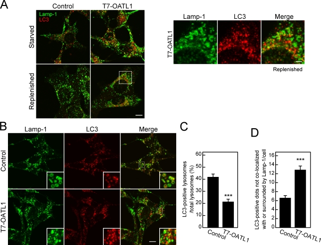
Overexpression of OATL1 inhibited the encounter between autophagosomes and lysosomes. (A) The residual LC3-positive structures did not contain Lamp-1. MEF cells stably expressing T7-OATL1 or not expressing T7-OATL1 (control) were cultured under starved conditions and replenished conditions. The cells cultured under each condition were fixed and stained with anti-LC3 antibody and anti–Lamp-1 antibody. Merged images are shown. Higher magnification views of the boxed area are shown on the right. (B–D) Overexpression of OATL1 caused a reduction in the ratio of LC3-positive lysosomes. The cells treated with bafilomycin A1 under starved conditions were fixed and stained with anti-LC3 antibody (red) and anti–Lamp-1 antibody (green). Merged images are shown on the right. Higher magnification views of the boxed areas are shown as insets. The ratios of LC3-positive lysosomes and the numbers of Lamp-1–negative LC3 dots per cell are shown in C and D, respectively. Error bars represent the means ± SEM of representative data (n ≥ 100) from two independent experiments. ***, P < 0.001; Student’s unpaired t test (compared with the control under the same conditions). Bars: (A [left] and B) 10 µm; (A, right) 2 µm.

