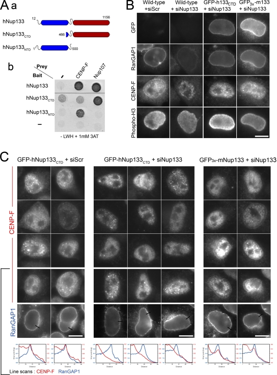
Figure 2.
The N-terminal domain of hNup133 tethers CENP-F at the NE in prophase. (Aa) Schematic representation of hNup133 constructs used in this study outlining its previously described β-propeller (N-terminal domain, NTD, blue) and α-solenoid (C-terminal domain, CTD, red) domains (adapted from Berke et al., 2004). (Ab) Yeast two-hybrid interactions between hNup133 (aa 12–1156), hNup133CTD (aa 466–1156) or hNup133NTD (aa 1–500), and hNup107 (aa 784–924), or CENP-F (aa 2644–3065) were analyzed as described in Materials and methods. Empty bait and prey vectors were used as negative controls (−). (B and C) Control HeLa cells (wild-type) or cell lines stably expressing GFP-hNup133CTD or GFP3x-mNup133 were transfected with a scramble siRNA (siScr) or with an siRNA duplex targeting the N-terminal domain of human Nup133. Cells were fixed after 3 d and processed for immunofluorescence using anti-RanGAP1, anti-CENP-F, and anti–phospho-H3 antibodies. Typical G2/M phospho-H3–positive cells are presented. Bars, 10 µm. In C, the anti-RanGAP1 signal is shown for cells on the bottom row. Line scans (with distances in pixels) measuring the intensity of CENP-F (red lines) and RanGAP1 (blue lines) reveal the peak of CENP-F that colocalizes with RanGAP1 at the NE in GFP-hNup133CTD treated with the scramble siRNA duplexes (left panels) or in GFP3x-mNup133 cells depleted for endogenous Nup133 (right panels), but not in Nup133-depleted GFP-hNup133CTD cells (middle panels). In some prophase cells, the bright intranuclear foci reflect the early recruitment of CENP-F at kinetochores.

