Figure 7.
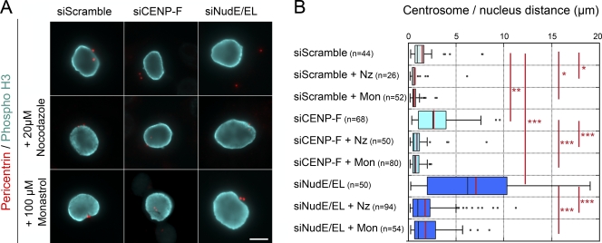
Centrosome movement away from the nuclear periphery requires microtubules and Eg5 activity. (A) HeLa cells transfected with scramble, CENP-F, or NudE/EL siRNA duplexes were either fixed (top row) or incubated with 20 µM nocodazole for 30 min or with 100 µM monastrol for 1 h before fixation. They were then stained with anti-pericentrin and anti–Phospho-H3 antibodies. Note that under those conditions, all phospho-H3–positive cells had entered prophase in the absence of microtubules or before Eg5 activation. All images arise from a single experimental dataset, although they were captured at different times using slightly different acquisition settings. Either a unique plane or maximum intensity projections of stacks are presented, as needed, depending on the locations of the centrosomes relative to the focal plane. Bar, 10 µm. (B) Distances between centrosomes and the NE, measured in phospho-H3–positive cells processed as above, are represented as box-plots using KaleidaGraph (see Materials and methods). The black and red bars indicate the median and mean values, respectively. The total number of cells quantified is indicated (n). ***, P < 10−5; **, P < 10−3; *, P < 0.05 obtained using the Student’s t test.

