Figure 3.
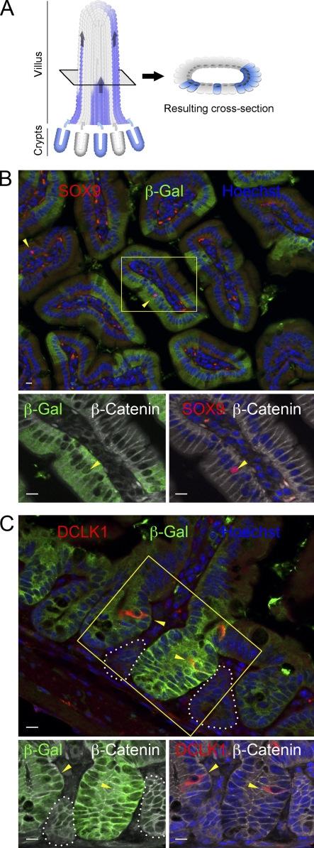
Tuft cells derive from Lgr5+ CBC stem cells. (A) Scheme explaining how chimeric Cre expression in crypts results in heterogeneous β-galactosidase staining in the adjacent villi (several crypts contribute to the generation of the cells constituting each villus). Wild-type (gray) and β-galactosidase (blue) cells coming from un-recombined (gray) and recombined (blue) crypts can migrate and colonize the same villus. The resulting cross section is shown. (B) Immunofluorescent staining for SOX9, β-galactosidase, β-catenin, and Hoechst in the Lgr5-EGFP-IRES-creERT2; Rosa26-LacZ mouse. Arrowheads point at SOX9+ tuft cells. The inset shows higher magnification of a SOX9+ tuft cells nucleus within a stretch of β-galactosidase+ cells. (C) Immunofluorescent staining for DCLK1, β-galactosidase, β-catenin, and Hoechst in intestinal sections from the Lgr5-EGFP-IRES-creERT2; Rosa26-LacZ mouse line. Arrowheads point at DCLK1+ tuft cells. The inset shows higher magnification of two DCLK1+ tuft cells within β-galactosidase+ crypts. β-galactosidase- crypts are shown by white dotted lines. Bars, 10 µm.

