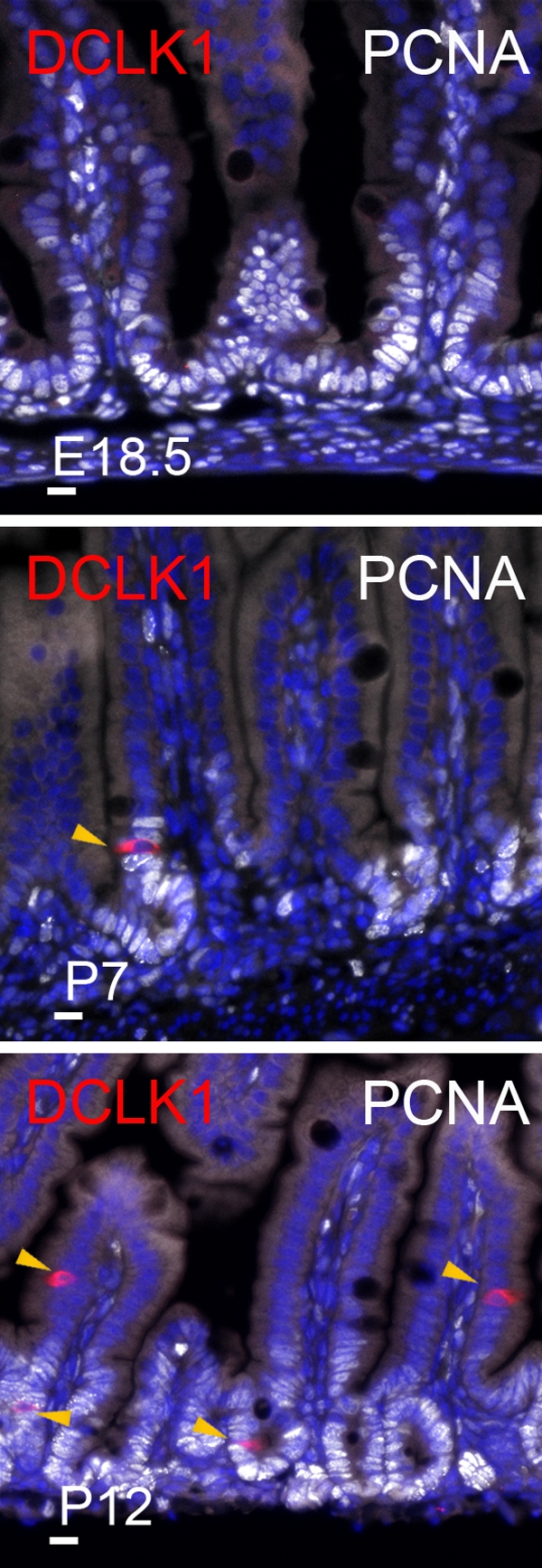
Figure 4.

Tuft cells appear after birth. Immunofluorescent staining for DCLK1 and PCNA in the developing small intestine of E18.5, P7, and P12 mice. Arrowheads point at DCLK1-expressing tuft cells. Nuclei are stained with Hoechst (blue). Bars, 10 µm.
