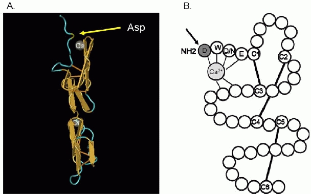
Figure 2.

Class I cbEGF domain showing the position of the first Asp in relation to the calcium molecule. (A) 3D picture of a cbEGF domain of fibrillin-1. The yellow arrow points to the first Asp. Picture derived from the NCBI database (http://www.ncbi.nlm.nih.gov/) (Downing et al., 1996). (B) cbEGD like domain (Handford et al., 1991). The arrow points to the first aspartic acid residue. Solid lines are the disulphide bridges between cysteine residues.
