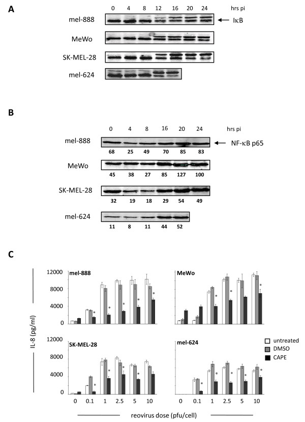
Figure 2.
Reovirus infection activates NF-κB in melanoma cells to induce cytokine secretion. (A) Melanoma cell lines were seeded in 100 mm dishes and treated with 10pfu/cell reovirus. At 4, 8, 12, 16, 20 and 24 hours post-infection whole cell lysates were prepared and I-κB assessed by western blot. (B) Melanoma cell lines were seeded as in (A), and nuclear fractions were prepared and western blotted for NF-κB p65. Densitometry data is shown underneath each blot. (C) Melanoma lines were seeded in 24 well plates and pre-treated with 50 μM CAPE, or equivalent DMSO solvent concentrations, for 2 hours prior to addition of reovirus at the indicated doses. Supernatants were collected after 48 hours and IL-8 levels determined using ELISA. Data are representative of at least 3 independent experiments. * indicates P < 0.05, by Student's t-test.

