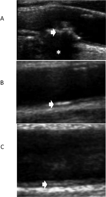
Figure 1.
Ultrasound exams of different types of VC. (A) Type V atheroma plaque (white arrow) with acoustic shadowing because of plaque calcification (asterisk). (B) Linear hyperechogenicity located in the lumen-intima interphase (white arrow). (C) Linear hyperechogenicity located in the media space (white arrow).

