Figure 1.
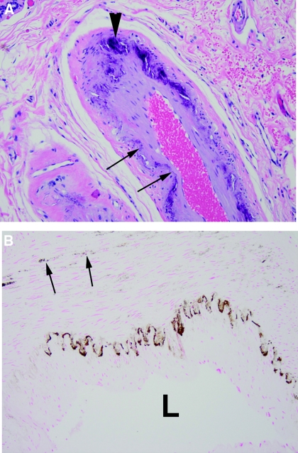
Histology of breast arterial calcification. (A) Hematoxylin and eosin stain showing linear calcification of the internal elastic lamina (arrows) and large deposits within the media (arrowheads). (B) von Kossa stain showing linear staining of the internal elastic lamina and granular staining within the media (arrows). L indicates the lumen.

