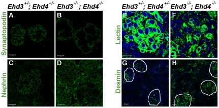
Figure 7. Alteration in endothelial, podocytic and mesangial cells in Ehd3–/–; Ehd4–/– glomeruli.
(A–D, G–H) Kidney sections from 39 day old Ehd3 +/–; Ehd4 +/– and Ehd3–/–; Ehd4–/– mice were immunostained with antibodies to synaptopodin (A–B), nephrin (C–D) and desmin (G–H) as described in Materials and Methods. (E–F) Endothelial cells were visualized by labeled tomato lectin staining (green) and DAPI (blue) was used to counterstain nuclei. Markedly altered staining patterns were observed in the Ehd3–/–; Ehd4–/– kidney sections in each case. Scale bar = 20 µm in panels A–F and 10 µm in panels G–H. White lines demarcate glomeruli in panels G–H.

