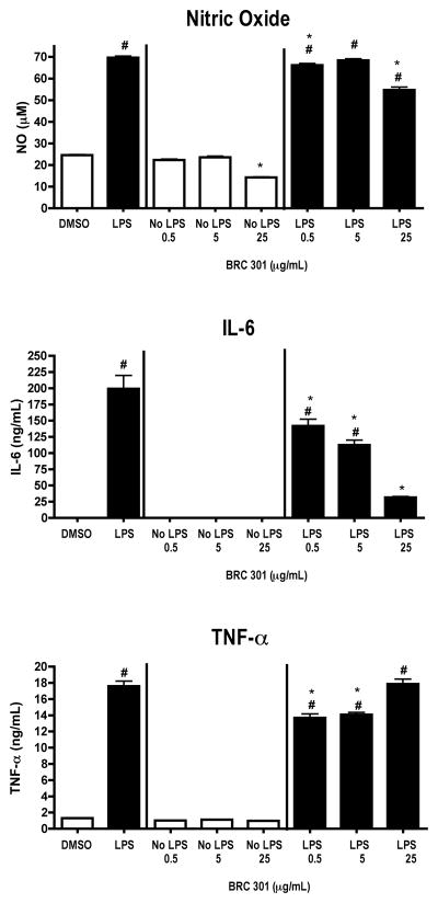
FIGURE 1.
LPS-activated DC2.4 cells produce decreased levels of NO, TNF-α, and IL-6 following exposure to BRC-301. DC2.4 cells were stimulated with LPS (1 μg/ml) and concomitantly treated with DMSO (0.1%) or BRC-301 (0.5, 5.0, 25 μg/ml) for 48 hours. Supernatants were collected and analyzed by ELISA (TNF-α and IL-6) or Griess Reagent System (NO). Results are mean ± SEM (n=3) and are representative of two separate experiments. # indicates p < .05 compared to the unstimulated control; * indicates p < .05 compared to the vehicle control.

