FIGURE 5.

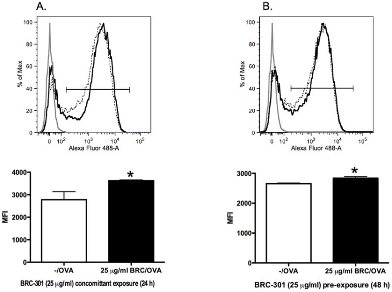
Phagocytosis of soluble antigen by bmDCs increases following exposure to BRC-301. BmDCs were treated with DMSO (0.1%) or BRC-301 (25 μg/ml) under the two following conditions. BRC-301 and Alexa Fluor 488-ovalbumin were added simultaneously and incubated overnight (A). BRC-301 was added 24 hours prior to exposure to Alexa Fluor 488-ovalbumin in pre-treated samples and cultured for an additional 24 hour incubation period (B). Cells were then harvested and analyzed by flow cytometry. Light gray lines represent control cells that received no antigen; dotted lines represent vehicle-treated bmDCs exposed to antigen; solid lines represent BRC-301-treated bmDCs exposed to antigen. Results are reported as Mean Fluorescence Intensity (MFI) ± SEM (n=3) and are representative of two separate experiments. * indicates p < .05 compared to the vehicle control.
