
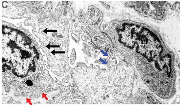
Figure 4. Endothelial cell activation.

Ultrastructural appearance (10,000x magnification, patient #2) of capillaries pre and post left ventricular assist device (LVAD) unloading revealed strong evidence of endothelial cell activation post LVAD. Panel A (pre LVAD). Red arrowheads: basal lamina, small blue arrows: cytoplasmic organelles and nuclei, big red arrow: capillary lumen.
Panel B (post LVAD). Red arrowheads: basal lamina reduplication, small blue arrows: increased nuclei size and increased cytoplasm organelles protruding into the capillary lumen- irregular luminal surface (big red arrow)
Panel C (post LVAD). Basal lamina reduplication (red arrows), increased nuclei and cytoplasmic size with increased pinocytotic vesicles protruding into the capillary lumen (black arrows), and numerous irregular lumenal and surface membrane projections (blue arrows), all indicative of endothelial activation.