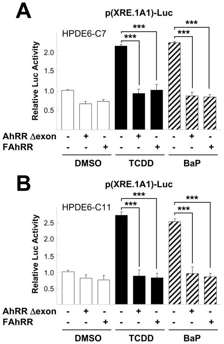
Fig. 4.
AhRR reduced TCDD or BaP-stimulated p(XRE.1A1)-Luc reporter activity. Pancreatic cells, HPDE6-C7 (A) and HPDE6-C11 (B), transfected with pCDNA3, AhRR Δexon, or FAhRR as well as p(XRE.1A1)-Luc for overnight were further incubated with 10 nM TCDD or 5 μM BaP for 24 hr then harvested to measure luciferase activity. All experiments were performed three times. The two-tailed Student t test was used for statistical analysis and the symbol (***, P < 0.001) in the figure indicates statistically significant differences.

