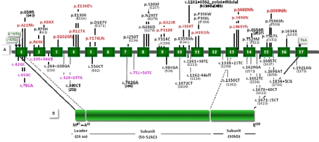
Figure 1.

Distribution of the detected mutations and polymorphisms in relation to the GALC gene and GALC polypeptide. In the top half of the diagram (A), the map of the GALC gene depicts the positions of the seventeen exons (dark green boxes numbered 1 to 17) and associated unnumbered introns (gray lines). In the bottom half of the diagram (B), the schematic representation of the polypeptide shows the 26-amino acid signal (leader) sequence, and the two protein subunits (50-52 kDa and 30 kDa) predicted to be proteolytically processed from the precursor protein. All mutations encountered in this patient series are given in (A) above the gene schema, all polymorphisms below. Novel mutations and polymorphisms reported here are denoted in red and violet respectively. HGVS nomenclature guidelines stipulate amino acid numbering from the first methionine of the 42-residue signal sequence. Hence, HGVS nomenclature was used for the mutations and the polymorphisms reported here (with the traditional designations given in parentheses).
