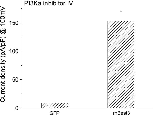
FIGURE 4.
PI3Kα inhibitor IV and Best3 activation. Average amplitudes of mBest3 currents activated by PI3Kα inhibitor IV. Wild-type mBest3 in pcDNA3.1 vector was cotransfected into HEK cells with GFP or cells were transfected with GFP only. Green fluorescent cells were selected for whole-cell recordings with ramp voltage stimulations (refer to Fig.1 legend). The PI3K inhibitor (10 μM) applied to the bath dramatically stimulated Cl currents from mBest3-transfected (n = 7) but not from GFP-only transfected HEK cells (n = 4). The currents obtained with 100-mV stimulation, 10 minutes after the PI3K inhibitor application to two groups, were averaged for statistical analysis (P < 0.01).

