Fig. 1.
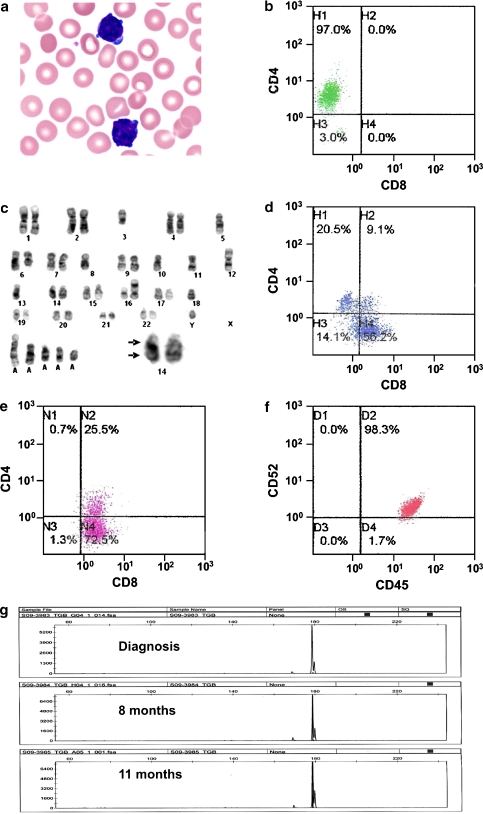
Pathological features of a case of T-cell prolymphocytic leukemia (PLL). a Abnormal lymphoid cells at diagnosis, showing condensed nuclei with inconspicuous nucleoli, and scanty basophilic cytoplasm with frequent blebs (Wright–Giemsa stain, original magnification ×100). b Flow cytometric analysis at diagnosis, showing predominance of CD4+ PLL cells. c Karotyping analysis, showing inv(14)(q11q32) (arrow, insert) and other aberrations. d At 8 months, there was an increase in CD8+ PLL cells. e At 11 months, there was predominance of CD8+ PLL cells. f CD52 expression had been consistent through the clinical course. g T cell receptor gene gamma rearrangement studies, showing identical peaks at various time points in the clinical course (GeneMapper v3.7, Applied Biosystems)

