
Fig. 3.
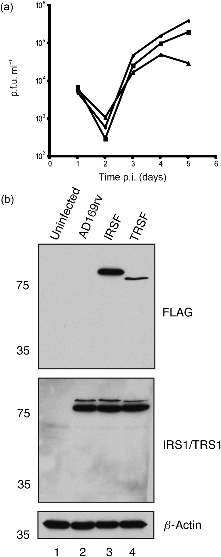
Characterization of recombinant virus. (a) Replication of AD169rv (•), IRSF (▪) and TRSF (▴) viruses. HFF cells were infected at an m.o.i. of 1 and virus supernatant was harvested at the indicated time points. Virus titre is represented as p.f.u. ml−1 on HFF cells. Data are representative of two experiments. (b) Western blotting of infected cells. Cell lysates of uninfected HFF cells (lane 1) or HFF cells infected at an m.o.i. of 1 with AD169rv (lane 2), IRSF (lane 3) or TRSF (lane 4) viruses were prepared 72 h p.i. Proteins in each lane were examined by Western blotting for the presence of FLAG-tagged IRS1 and FLAG-tagged TRS1 (top panel), IRS1 and TRS1 (middle panel) and β-actin (bottom panel) using antibodies recognizing these proteins or FLAG, as indicated to the right of the figure. Note that, in the middle panel, a combination of antibodies recognizing IRS1 or TRS1 was utilized and that FLAG-tagged versions of each protein migrate more slowly than wild-type proteins. The positions of molecular mass markers (in kDa) are indicated to the left of the figure.