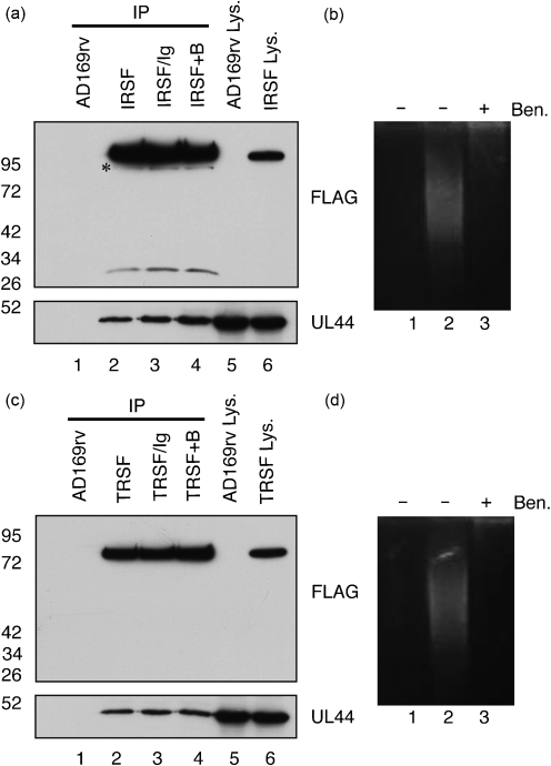
Fig. 4.
Detection of protein immunoprecipitated from IRSF- and TRSF-infected cell lysate by Western blotting. (a, c) Proteins immunoprecipitated (IP) from lysate of AD169rv- and IRSF-infected cells (a) or AD169rv- and TRSF-infected cells (c) prepared 72 h p.i. were separated on a 10 % polyacrylamide gel and examined by Western blotting using antibodies recognizing FLAG (top panels) or UL44 (bottom panels). Immunoprecipitated proteins are examined in lanes 1–4. Lysates precleared with control antibody before IP (IRSF/Ig or TRSF/Ig) are shown in lane 3 of both figures. Lysates treated with Benzonase (IRS+B or TRS+B) are shown in lane 4 of both figures. Lysates (Lys.) used in the IP are examined in lanes 5 and 6. The positions of molecular mass markers (in kDa) are indicated to the left of the figure. The novel IRS1 band discussed in the text is marked with an asterisk. (b, d) Cell lysate used in the IP in lanes 2 and 4 of (a) and (c) was run out on an ethidium bromide-stained 0.8 % agarose gel [(b) and (d), respectively]. Lanes: 1, no sample; 2, IP in the absence of Benzonase (Ben.); 3, IP in the presence of Ben.

